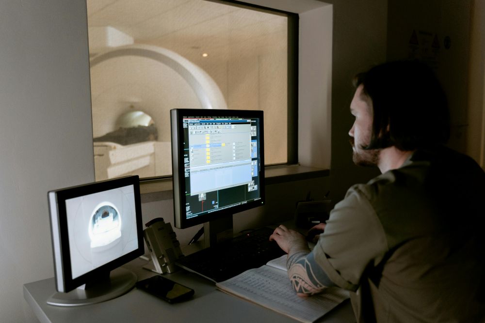
The new method helps to map skin cancer more clearly before surgery.
A team of scientists may have found a new way to help doctors see and treat skin cancer more effectively. This approach uses a special type of imaging that can create a clear, three -dimensional images of the tumor inside the skin. The procedure combines light and sound so that it can produce a detailed picture of what is happening under the surface. This helps doctors see the actual shape and depth of the tumor, which needs to be cut off without the first skin. He tested it on the most common type of skin cancer patients, called basal cell carcinoma, or briefly BCC.
Basel cell carcinoma is like skin cancer that many people get, especially when they grow older. In Singapore, matters are increasing, and older adults are watching it more often. Generally, when there is a suspicious place on one’s skin, it requires biopsy to know if it is cancer or not. This means that a small piece of skin is removed and checked under a microscope. If it is a cancer, Dr. Mohaus can remove the tumor using a method called surgery, where he cut the skin slightly and check each piece under microscope until cancer is found. Although it is effective, it may take a lot of time and can be restless. Some people also need more than one surgery if the first tumor was not removed.

The new technique hopes to change it all. Singapore’s Agency for Science, Technology and Research Scientists have worked together with doctors at the National Skin Center to test a new machine, which uses something called multi -spectrolous optoccostic tomography. It’s a mouth, but the idea is easy. The machine sends light to the skin, which causes a little heat. This heat creates sound waves that bounce backs and help create a picture. After that, an automatic computer program offers a tumor shape. The system shows how wide, deep and tall the tumor is – such as making a 3D map inside the body. This helps the doctor to see how much it needs to be removed, which makes the surgery faster and more precise.
In the first test of this technology, eight patients scan their tumors before surgery. The scans regularly match the consequences of doctors after surgery using regular methods. It is a good sign that the machine is doing its job well. It also shows that this system can help prevent additional surgeries for the first time. Low surgery means low pain and low maintenance for patients.
Although this new imaging tool was made for basal cell carcinoma, Singapore researchers believe it can help with other types of skin cancer. The team is still examining it, but they hope it can start something bigger. If this system continues to perform well, doctors to treat skin cancer can soon have a new way that is fast, easy and more accurate.
For now, the team will test and improve the system. They want to ensure that it works well in different types of skin and in all kinds. But if everything goes well, it can be part of regular cancer care, which gives patients a smooth way for treatment and recovery.
Sources:
-No 3D imaging technique increases the diagnosis of basal cell carcinoma
A proof off -conceptual study for precise mapping of color basal cell carcinoma in Asian skin using Level Set Segency Imaging
